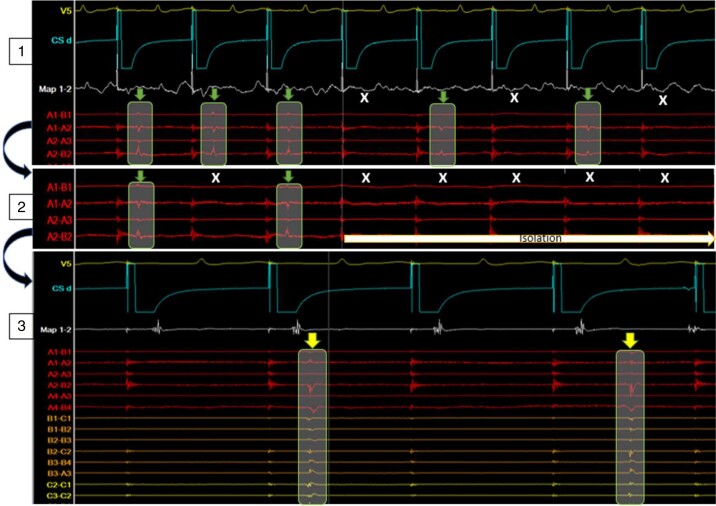
Figure 4.
Intracardiac EGM during anterior–posterior approach: HD grid on posterior wall, CS catheter pacing from RAA, and ablation catheter anterior to RSPV as indicated in Figure 3. Top (1) Ablation energy delivery at 12.18 p.m. causing 2:1 block (green arrows = conduction, white X = block) into the box as recorded by the HD grid. Middle (2) with continuation of energy delivery after eight beats of 2:1 conduction successful isolation of the posterior wall was achieved. Bottom (3) at 12:35 p.m., 17 min after isolation, ongoing independent activity within the box as recorded by the HD grid (yellow arrows). CS, coronary sinus; EGM, electrogram.

