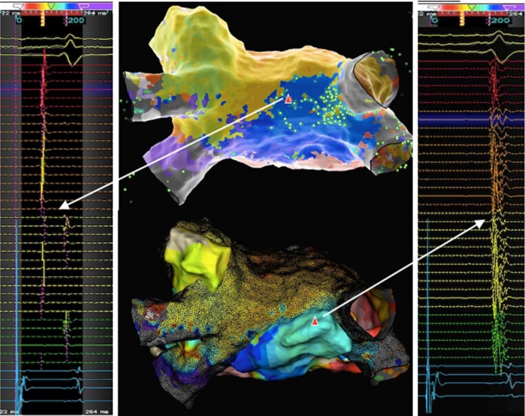
Figure 6.
Case vignette endo-epicardial AF ablation: 43-year-old patient with symptomatic persistent atrial fibrillation (AF) with severely impaired left ventricular function secondary to uncontrolled rate and previous PVI and posterior box isolation. Mapping with an Advisor™ HD grid during CS pacing confirmed blocked floor but connected roof line. View on left atrial roof. Top and left: endocardial map with evidence of widely split double potentials (∼100 ms) on HD grid splines positioned along roof line. Note, earliest activation of posterior wall occurs at right superior corner at site of insertion of septo-pulmonary bundle (highlighted by “sparkles”, green bright dots). Bottom and right: Epicardial map (full colour) superimposed on endocardial map (black mesh) with evidence of long fractionated signals on opposing site to the earliest endocardial activation of the posterior box (adapted from Tonko et al.42). Ablation at the corresponding endo- and epicardial sites of suspected connection over the roof successfully isolated the posterior wall with no reconnection after a waiting time of 30 min. AF, atrial fibrillation; CS, coronary sinus; PVI, pulmonary vein isolation.

