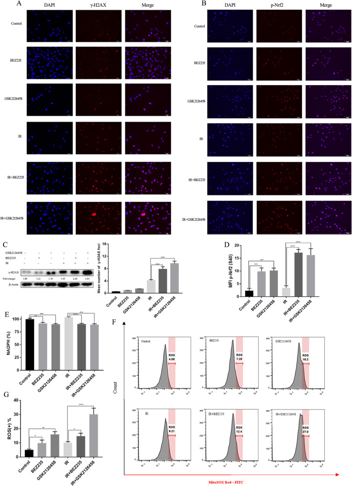
Fig. 5. PI3K/mTOR inhibitors intensified DNA damage and oxidative damage caused by IR in H446RR cells.
A γ-H2AX expression across various treatments were determined by immunofluorescence. B The expression of p-Nfr2 (S40) across different treatments were determined by immunofluorescence. C γ-H2AX level across various treatments was quantitatively analyzed, and γ-H2AX expression were detected by immunoblotting. D The p-Nfr2 (S40) expression across different groups was quantitatively analyzed. E NADPH level in cells treated with PI3K/mTOR inhibitors combined with or without IR was detected. F, G ROS level of H446RR cells across different groups was detected using MitoSOX Red, and the mean level of ROS were obtained.

