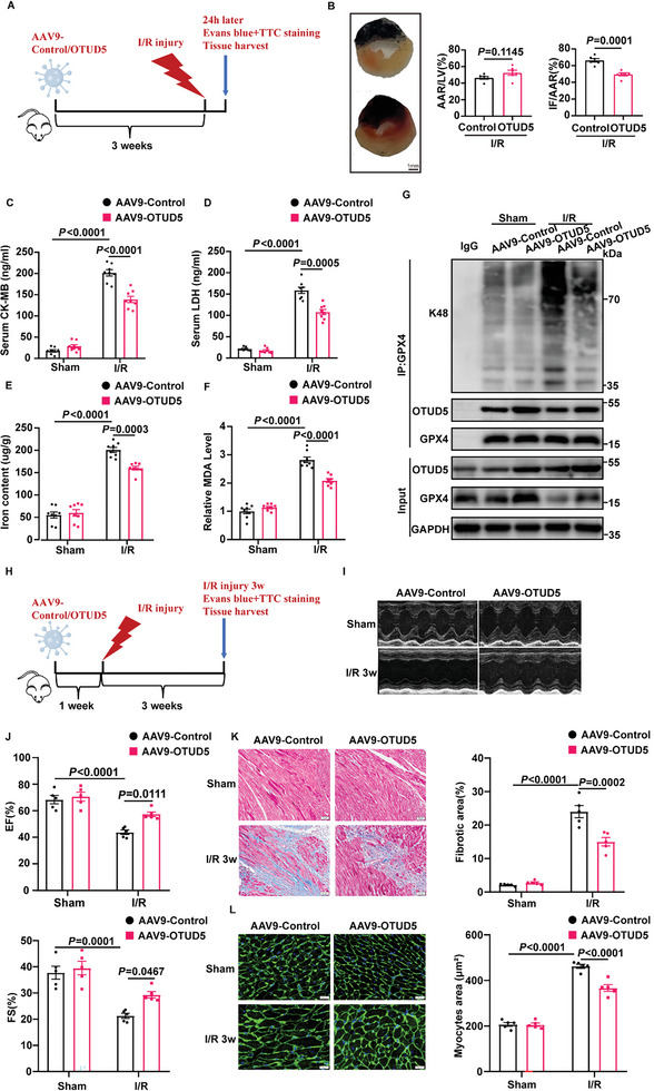
Figure 7.

OTUD5 overexpression prevents cardiac ischemia/reperfusion injury in ALDH2 cKO mice. A) Schematic diagram showing that AAV9‐Control or AAV9‐OTUD5 were injected via tail vein, and 3 weeks later mice were subjected to cardiac ischemia/reperfusion (I/R) injury for 24 h. B) Representative sections and quantitative data for infarct size (IF) and area at risk (AAR) in mice hearts subjected to I/R injury (30‐min ischemia/24‐h reperfusion, n = 6). Scale bar: 1 mm. C,D) Serum levels of CK‐MB and LDH in mice with sham or MI/R injury (30‐min ischemia/24‐h reperfusion, n = 8). E,F) Iron content and MDA levels in mice subjected to MI/R (30‐min ischemia/24‐h reperfusion, n = 8). G) The ubiquitination of GPX4 was determined by immunoprecipitation with anti‐GPX4 antibody in mice subjected to MI/R injury (30‐min ischemia/24‐h reperfusion, n = 4). H) Schematic diagram showing that AAV9‐Control or AAV9‐OTUD5 were injected via tail vein, and 1 week later mice were subjected to cardiac ischemia/reperfusion (I/R) injury for 3 weeks. I,J) Echocardiography for left ventricular ejection fraction (EF, %) and fractional shortening (FS, %) in mice after I/R injury (30‐min ischemia/3‐week reperfusion, n = 5). K,L) Masson Trichrome staining for cardiac fibrosis (Scale bar: 50 µm) and WGA staining for cardiac hypertrophy (Scale bar: 20 µm) in heart tissues of mice after MI/R (30‐min ischemia/3‐week reperfusion, n = 5). Data are expressed as mean ± SEM. Unpaired two‐tailed Student's t‐test was used for the analysis in B). One‐way ANOVA was used for the analysis in C–F,J–L).
