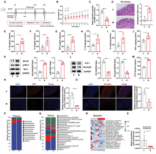
Figure 1.

Rectal administration of AA induced PI‐IBS and caused gut microbiota disorder in rats. A) Experimental scheme of the PI‐IBS rat model used in this study (n = 5). B) Changes in body weight of PI‐IBS rats (n = 5). C) Water injection rate when the AWR score was 3 (n = 5). D) H&E staining and pathological score of colon sections (n = 3). E–J) Concentrations of the representative proinflammatory cytokines IL‐1β, IL‐6, and TNF‐α in rat serum samples of colon tissue (n = 4‐5). K) Expression of key proteins of the MLCK/p‐MLC signaling pathway in rat colon tissue samples (n = 4). L) The expression of TJ proteins (ZO‐1, Occludin) in rat colon tissue (n = 4). M) Measurement of gut permeability in rats (n = 3). N and O) Immunofluorescence staining of TJ protein (ZO‐1, Occludin) expression in the colonic section (n = 3). Q,R) Relative abundance of the identified fecal microbiota at the (P) phylum, (Q) genus, and (R) species levels as per 16S rRNA gene sequencing (n = 5). S) The relative abundance of B. acidifaciens. C, control group; M, PI‐IBS rat group. Scale bar, 100 µm. Data are presented as the mean ± SD. ns, p > 0.05; * p < 0.05; ** p < 0.01; *** p < 0.001. AA: Acetic acid; PI‐IBS: Post‐infectious irritable bowel syndrome; AWR: Abdominal withdrawal reflex; TJ: Tight junction; PCoA: Principal coordinate analysis.
