Figure 1:

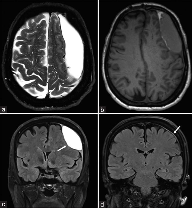
Illustrative case no. 1 of interdural hematoma. (a) Preoperative T2-weighted axial magnetic resonance imaging (MRI); (b) preoperative T1-weighted axial MRI; (c) preoperative T2 fluid attenuated inversion recovery (FLAIR) coronal MRI showing the biconvex lentiform hematoma (white arrow); and (d) 6 months postoperative T2 FLAIR coronal MRI showing the complete resolution of the collection with a slight residual dural thickening (white arrow). Notice the irregular thick inner wall of the hematoma (a). Notice the dural thickening at both dural edges (dural beak sign) indicating dural splitting and the slight subarachnoid space enlargement (c).
