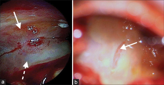
Figure 2:

Intracranial, interdural endoscopic pictures taken through a single burr-hole, after hematoma evacuation from case no. 1. (a) Image showing the dural splitting from the inside of the hematoma pocket at the dorsomedial apex (dashed white arrow points to the inner dural layer and solid white arrow to the outer dural layer). (b) Image of the inner wall after dural biopsy, revealing the transparent thin arachnoid membrane (solid white arrow).
