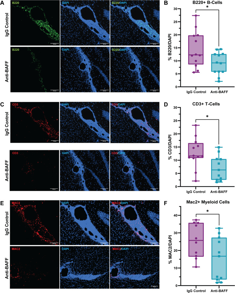
Fig. 2.
Anti-BAFF antibody 10F4 reduced leptomeningeal inflammation detected through histopathology. A, B On pathological evaluation at Week 10, we observed a decrease in the proportion of B cells within the meningeal infiltrate in the anti-BAFF antibody 10F4 group compared to the IgG control group. C, D Additionally, the treatment with the anti-BAFF antibody 10F4 led to a significant reduction in CD3 + T cells compared to control IgG-treated EAE mice. E, F We also found a significant difference in the proportion of myeloid cells (Mac2 +) between the anti-BAFF antibody 10F4 and IgG control group, with Mac2 + cells being less in the anti-BAFF treatment group. The number of EAE mice for the anti-BAFF antibody 10F4 group was 12 and for the IgG control group was 12. For B, D, F, data are shown as box plots, the center line indicates the median, the box indicates the 25th and 75th percentiles, the whiskers range, and the dots indicate all data points. The statistical analysis was conducted using two sample Welch’s t-test as data were normally distributed. *P < 0.05. Scale bars = 100 um

