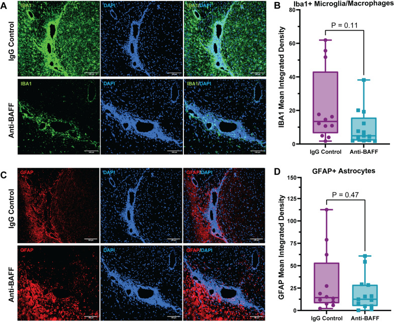
Fig. 3.
Effects of anti-BAFF antibody 10F4 on glia in adjacent brain tissue. A, B Histopathological immunofluorescence evaluation of IBA1 immunoreactivity revealed no significant change in the immunoreactivity of IBA1 in the cortex adjacent to the areas of meningeal inflammation in the anti-BAFF treatment group compared to the IgG control group. C, D No significant changes were observed in astrocytosis detected by GFAP immunoreactivity in the cortex surrounding the regions of meningeal inflammation. B, D Data were presented as box plots with the center line indicating the median, the box indicating the 25th and 75th percentiles, the whiskers indicating range, (min–max) and the dots representing all data points. The statistical analysis was conducted using Mann–Whitney U-tests, and P-values were derived from it. The significance level was set to *P < 0.05. The scale bars = 100 um

