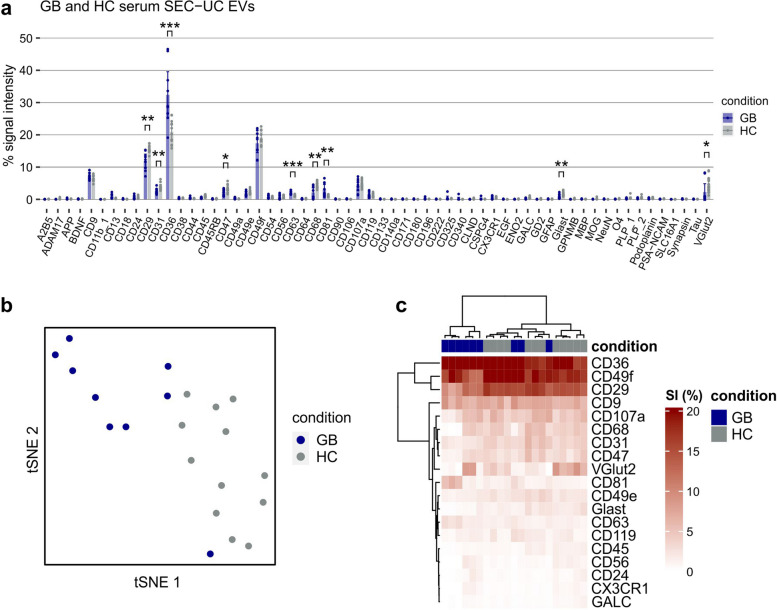
Fig. 4.
Profiling of serum EVs from GB patients and healthy controls. a Relative EV Neuro signal intensities for EVs separated from the serum of GB patients or HC by SEC followed by UC calculated as signal of target divided by the total signal of all markers (in %). Bars represent mean values and error bars indicate the 95% confidence interval. Asterisks mark statistically significant differences between GB and HC. Only significant alterations of targets that were consistently detected above background in all individuals are marked. *=p < .05, **=p < .01, ***=p < .001. (b) tSNE on data from (a) stratified by condition (color). c Heatmap visualization of selected targets from (a) including hierarchical clustering for targets as well as subjects

