
Fig. 8.
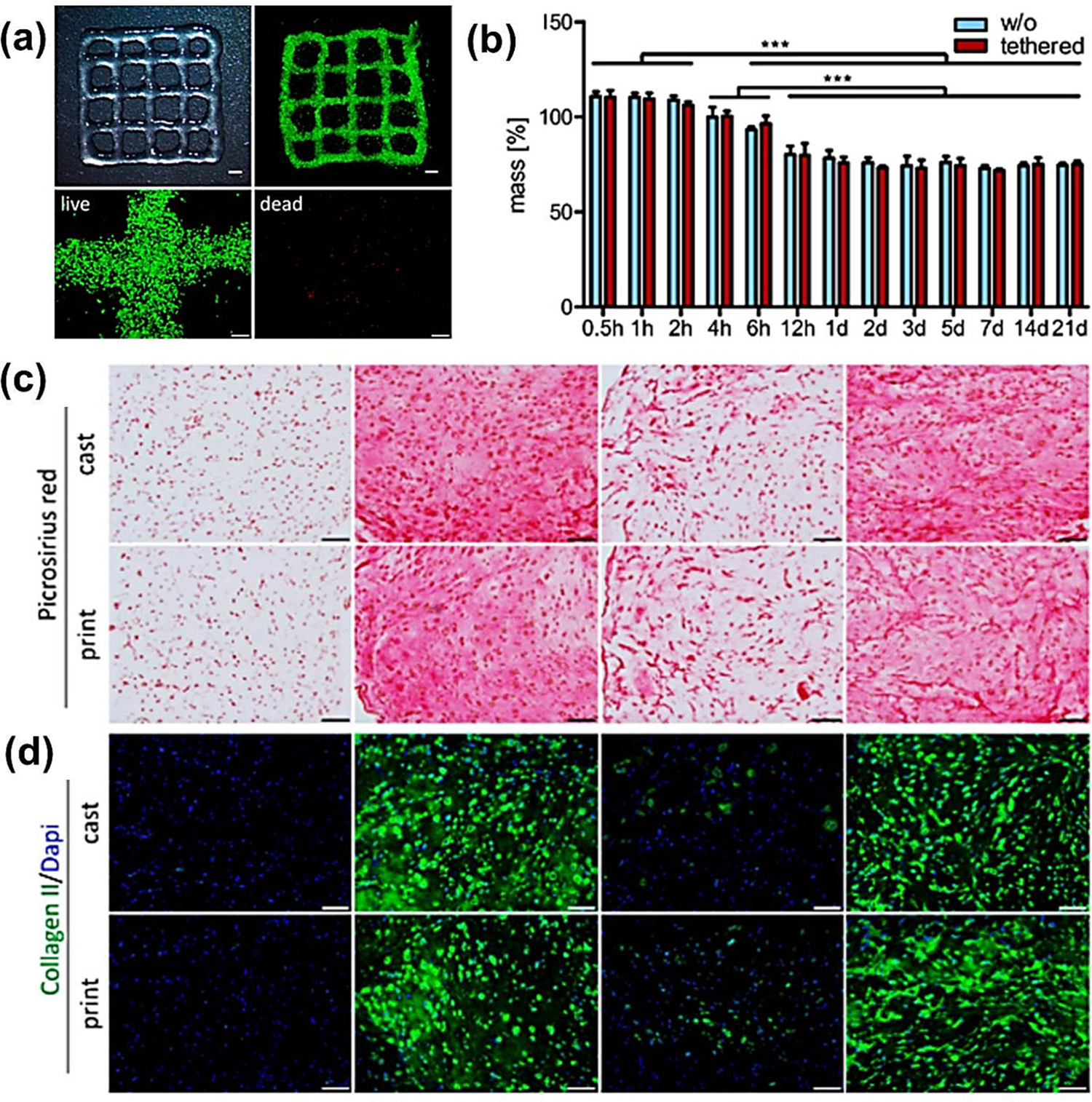
3D printing and bio-ink characterization (a) a 3D printed HA bio-ink grid (scale 2 mm), live (calcein-AM - green) dead (EthD-III – red) staining of the zoomed grid (scale 200 μm); (b) three week (Histological and immunohistochemical (IHC) staining based) swelling analysis (with and without) tethered TGF-β1. [Mean ± standard deviation (n = 3), significancy *** (p < 0.001)]. (c) Picrosirius red staining and (d) IHC staining for collagen type II. Nuclei - DAPI (Scale 100 μm) [124].