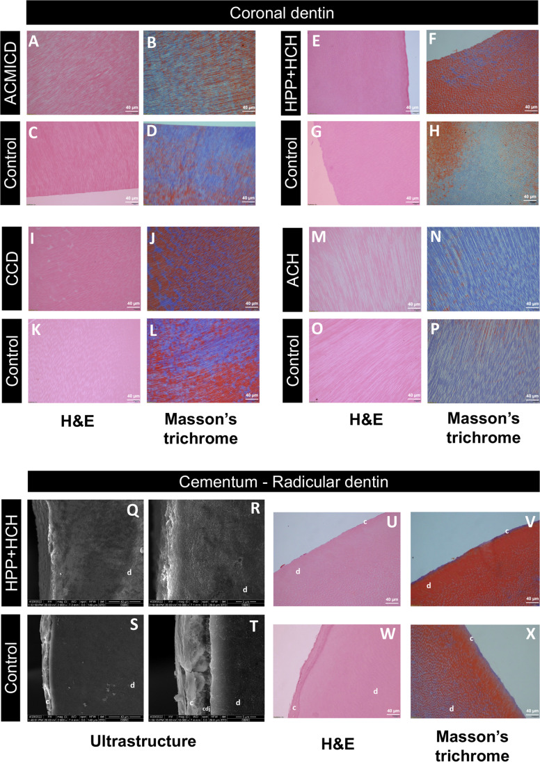
Fig. 4.
Histological analysis. (A–P) SD teeth had a dentinal tubule arrangement comparable to controls. (Q–X) A well-defined cementum layer and cementodentinal junction (CDJ) were observed in control roots (S, T, W, X), but not in HPP + HCH roots (Q, R, U V). Hematoxylin and eosin staining (× 20): A, C, E, G, I, K, M, O, U, W; Masson’s trichrome (× 20): B, D, F, H, J, L, N, P, V, X; SEM of cementum and radicular dentin: Q-T. d, dentin; c, cementum

