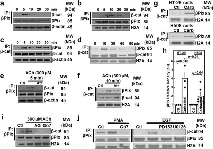
Figure 3.
MR activation stimulates βPix binding to β-catenin. (a-f) At the times shown, HT-29 cells were treated with 300 μM ACh. Cytosolic (a, c) and nuclear (b, d) fractions were probed after IP. (e–f) Pre-incubating cells with atropine (Atr, 5 μM for 30 min) for the indicated times blocked ACh effects in both the cytosol (e) and the nucleus (f). Ctl, control; β-actin and histone 2A (H2A) were used as cytosolic and nuclear fraction loading controls. (g) HT-29 and H508 human colon cancer cells were treated with 100 µM carbamylcholine (carb). Nuclear fractions were probed for βPix after IP; histone 2A (H2A) is a loading control. (h) Relative density (au, arbitrary units) was measured in six different βPix immunoblots performed as illustrated in (g), normalized to the loading control, and expressed as a function of βPix expression in unstimulated cells. N = 3 and 6 experiments for HT-29 and H508 cells, respectively. Ctl, control. Histone 2A (H2A) was a nuclear loading control. (i) HT-29 cells were pre-incubated with inhibitors of EGFR (AG1478) and PKCα/β1 (Gö6976) for 60 min before adding 300 µM ACh for an additional 10 min. After treatment, nuclear fractions were immunoprecipitated with anti-βPix antibody followed by immunoblotting as indicated. Histone 2A (H2A) was a nuclear marker. (j) In the left panel, HT-29 cells were treated with 50 nM PMA for 10 min with or without pre-incubation for 45 min with a PKC inhibitor (5 μM Gö6976). In the right panel, HT-29 cells were incubated with 10 ng/ml EGF for 10 min with or without preincubation for 60 min with EGFR (5 µM PD153035) and MEK (10 µM U0126) inhibitors. Nuclear fractions were probed by immunoprecipitation with anti-βPix antibody and immunoblotting with anti-β-catenin and anti-βPix antibodies. MW, molecular weight. Original uncut immunoblots are shown in Supplemental Materials.

