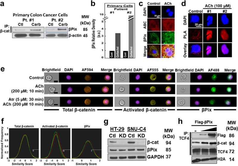
Figure 4.
MR agonist treatment of colon cancer cells induces co-localized cytoplasmic and nuclear βPix and β-catenin. (a) MR agonist stimulates βPix binding to β-catenin in primary colon cancer cells. Cells were treated with 100 µM carbamylcholine (carbachol; 10 min, 37 °C). Cytosolic fractions were immunoprecipitated with anti-β-catenin antibody, then immunoblotted with anti-βPix antibody. β-actin was a loading control. (b) Relative βPix signal intensity in extracts from vehicle- and carbachol-treated primary colon cancer cells. au, arbitrary units. (c) MR agonist-induced βPix and β-catenin co-localization. Primary colon cancer cells were treated with vehicle or ACh (100 µM, 10 min), then stained with DAPI (blue), anti-activated β-catenin (ABC, red), and anti-βPix (green) antibodies. In the merged images, yellow in the cytoplasm and purple in the nucleus undergoing mitosis (arrows) reveal co-localized βPix and β-catenin. (d) Proximity ligand assay (PLA) reveals MR agonist-induced nuclear βPix/β-catenin co-localization. Images show DAPI- and PLA probe (red)-stained primary colon cancer cells. Left, PLA reveals co-localized βPix and β-catenin in the cytoplasm of untreated cell (control; red dots). In primary colon cancer cells (middle and right), ACh stimulated nuclear co-localization of βPix and β-catenin (purple in overlay). (e) Dual-color images are shown after using flow cytometry to view HT-29 cells stained with fluorescence-tagged antibodies and nuclear dye [nucleus (DAPI), total β-catenin (AF594), activated β-catenin (AF555), and βPix (AF488)]. βPix nuclear translocation triggered by ACh (200 µM, 10 min) was blocked by pretreating cells with atropine (5 µM, 30 min). Left to right: brightfield, nucleus (blue), and, respectively, total β-catenin (brown), activated β-catenin (yellow), βPix (green), and merged images are shown. Scale bars, 10 µm. (f) Fluorescent cell sorting. Similarity scores for stained HT-29 cells are shown [control, green; ACh treatment, red; atropine pre-treatment then ACh, yellow]. (g) β-catenin knockdown does not alter βPix expression. HT-29 and SNU-C4 cells were transfected with β-catenin siRNA. Extracts were immunoblotted for β-catenin, βPix, and GAPDH (used as a loading control). Results shown are representative of three experiments. (h) βPix overexpression enhances nuclear β-catenin binding to TCF4. HCT116 cells were transfected with Flag-βPix and nuclear extracts were immunoprecipitated with anti-TCF4 antibody and immunoblotted for Flag and β-catenin. Histone 2A (H2A) was used as a loading control. MW, molecular weight. Original uncut immunoblots for (a, g), and h are shown in Supplemental Materials.

