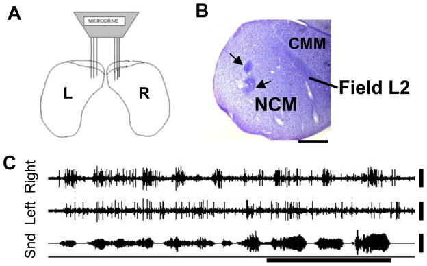
Figure 3.
NCM anatomy and electrophysiology. A) Schematic in coronal view of bilateral electrode placement in NCM. B) Sagittal section of NCM stained with cresyl violet. Black arrows indicate location of 2 electrolytic lesions made in NCM at the conclusion of recording. Scale bar: 1mm. Labels show locations of NCM, CMM and the primary auditory area, Field L2. C) Raw electrophysiological recording from electrodes placed in left and right NCM showing bilateral responses to conspecific song. X-axis time bar: 0.5s. Y-axis scale bar: 25μV

