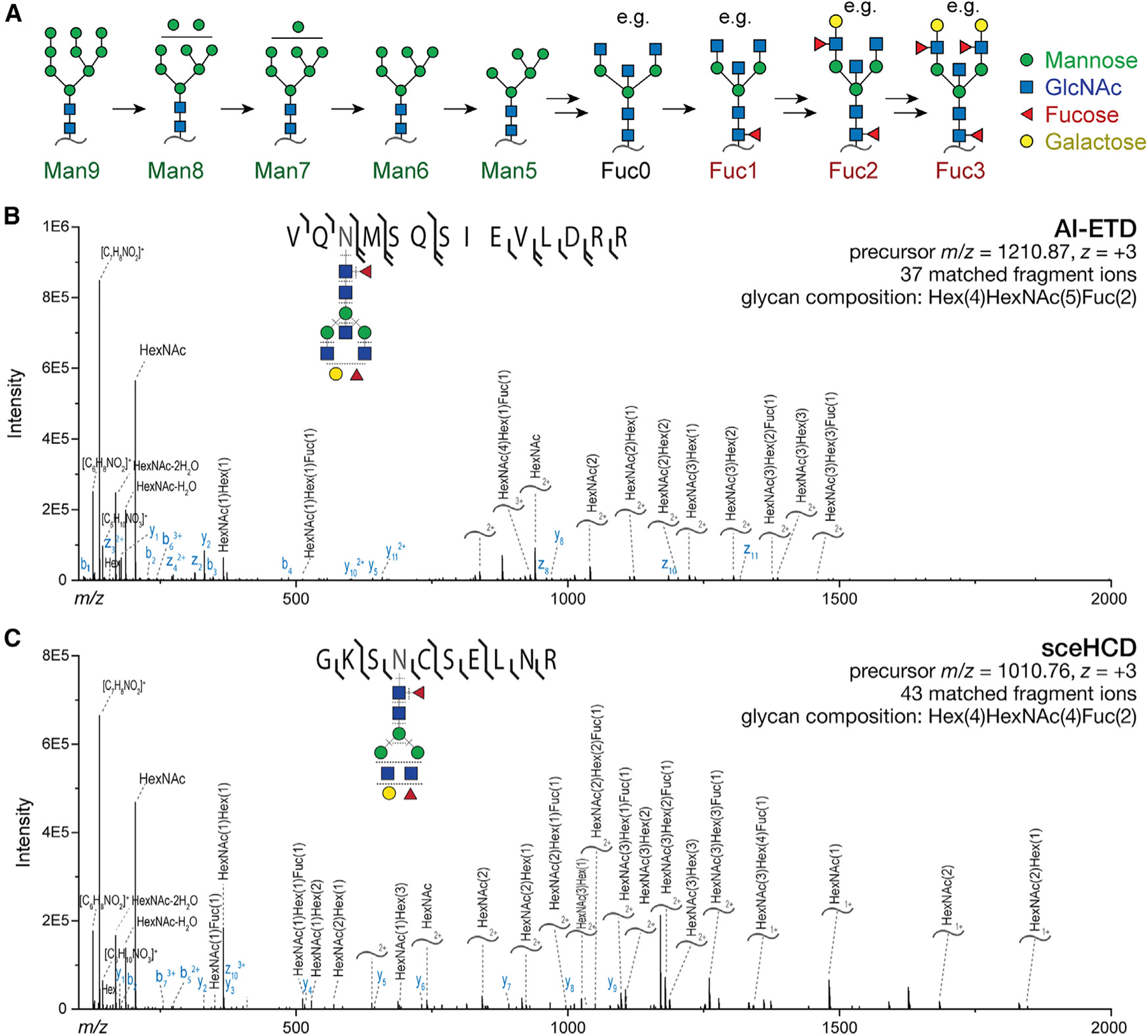
Figure 3. Characterization of protein N-glycosylation by MS/MS.

(A) Illustration of the N-glycan maturation sequence for the predominant brain N-glycans. Many potential glycans contain 0–3 fucoses, with some examples shown here.
(B) Illustrative annotated AI-ETD spectrum with glycopeptide precursor shown. Curved lines annotated with glycan compositions denote Y ions, which contain the intact peptide and a fragmented glycan. Both b/y and c/z ions are detected owing to hybrid vibrational and electron transfer modes of fragmentation.
(C) As in (B) but with sceHCD fragmentation of the depicted glycopeptide, which yields b/y and Y ions.
