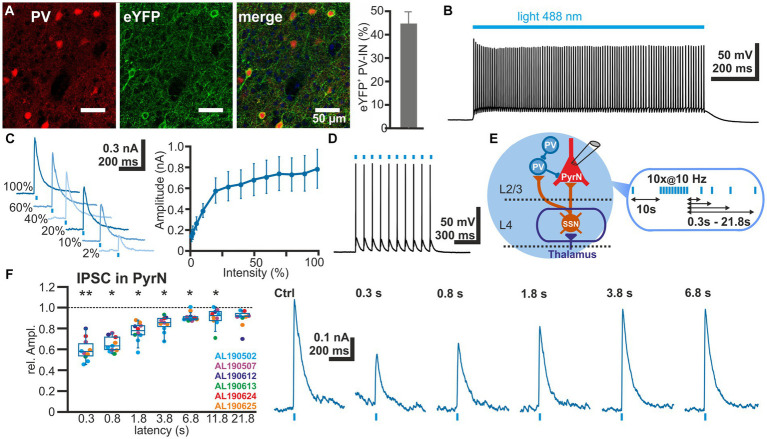
Figure 4.
Short-term plasticity of GABAergic synapses between PV interneurons and L4 pyramidal neurons. (A) Immunhistochemical staining of PV (left image) and of ChR2-associated eYFP expression (middle image) in slices of transfected animals. Note the exclusive eYFP expression in PV+ neurons and that 44.7 ± 4.9% (n = 20 ROIs) of the PV+ interneurons are transfected (right bar diagram). (B) Illumination with a 488 nm light pulse induced a persistent depolarization in an eYFP+ neuron. (C) The light-induced GABAergic IPSCs (determined at 0 mV) in L2/3 PyrN depend on the laser intensity. Already at 2% of the maximal laser intensity, substantial inward currents were induced, which saturated at >30% laser intensity. (D) The light-induced neuronal activity in PV interneurons can reliably follow 10 Hz optogenetic stimulation. (E) Schematic representation of recording and stimulation condition used for the experiment shown in panel (F). The stimulation protocol consisted of an optogenetic control stimulus, followed by optogenetic burst stimulation and optogenetic test stimuli applied between 0.3 and 21.8 s after the burst. GABAergic IPSCs were recorded in a L2/3 pyramidal neuron upon application of a stimulation protocol. (F) Boxplot illustrating that the relative amplitude of the optogenetically induced GABAergic IPSCs revealed a significant STD, lasting ca. 3.8 s. In the right panels typical current traces are displayed. Significance in panel (F) is indicated by *p < 0.05 and **p < 0.01.

