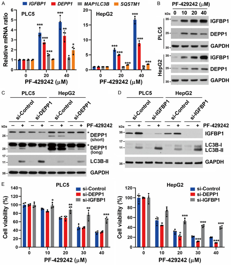
Figure 4.

Roles of IGFBP1 and DEPP1 in PF-429242-induced autophagic cell death in hepatocellular carcinoma (HCC) cells. A. PLC5 and HepG2 cells were treated with 20 and 40 μM for PF-429242 for 24 h, and then mRNA levels of the IGFBP1, DEPP1 (C10orf10), MAP1LC3B, and SQSTM1 (p62) genes were determined by a real-time qPCR. Error bars are the mean ± SD (n = 3). *, **, and *** indicate a significant difference (P < 0.05, P < 0.01, and P < 0.001, respectively) compared to untreated control cells. B. PLC5 and HepG2 cells were treated with 10, 20, and 40 μM PF-429242 for 24 h. Protein expressions were determined by Western blotting. C. PLC5 and HepG2 cells were transfected with siRNA specific for DEPP1 for 24 h, then treated with 20 μM PF-429242 for another 24 h. Protein expressions were determined by Western blotting. For DEPP1 expression, both short- and long-exposure images are shown. An asterisk (*) indicates non-specific bands. D. PLC5 and HepG2 cells were transfected with siRNA specific for IGFBP1 for 24 h, then treated with 20 μM PF-429242 for another 24 h. Protein expressions were determined by Western blotting. E. si-DEPP1/si-IGFBP1-transfected PLC5 and HepG2 cells were seeded in a 96-well plate and treated with the indicated doses of PF-429242 for 72 h. Cell viability was determined by Alamar Blue staining. Error bars are the mean ± SD (n = 4). *, **, and *** indicate a significant difference (P < 0.05, P < 0.01, and P < 0.001, respectively) between si-DEPP1/si-IGFBP1- and si-Control-transfected cells.
