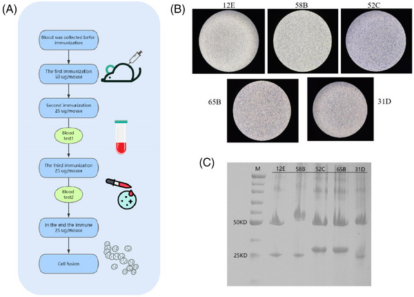
FIGURE 2.

Pairing of monoclonal antibodies. (A) Procedure for immunizing mice. (B) Microscopic image of five hybridoma cells, all of which are semi—adherent, semi—suspended cells. (C) SDS‒PAGE analysis of the purified antibody revealed that the heavy and light chains of the IgG antibody were visible at approximately 50 and 25 kDa, respectively.
