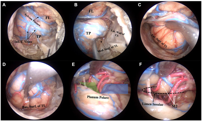
Figure 1.
Anatomic pictures showing a right endoscopic transorbital approach for the exposure and resection of the temporal lobe. Step 1 and 2, corresponding to the exposure of the temporal pole and the four surfaces of the temporal lobe, are shown. After dural opening and application of tucking sutures, the temporal pole, with its vascular network, comes into view (A). The mesial surface of the temporal lobe faces the lateral wall of the cavernous sinus inferiorly and the tentorial incisura superiorly (B). The lateral surface of the temporal lobe is detached from the lateral wall of the middle cranial fossa (C). After extradural flattening of the floor of the middle cranial fossa, the intradural exposure of the basal surface of the temporal lobe can be achieved (D). The sylvian fissure is split to expose the superior surface of the temporal lobe, the sphenoid (shaded blue area) and operculoinsular (shaded green area) compartments of the sylvian cistern, and the branches of the middle cerebral artery (E). The genu of the middle cerebral artery, with the M2 branches turning around the limen insulae, which sits at the lateral edge of the sphenoid compartment of the sylvian fissure, can be visualized (F). Bas. Surf. of TL, basal surface of the temporal lobe; FL, frontal lobe; Inf. T. Veins, inferior temporal veins; Lat. Surf. Of TL, lateral surface of the temporal lobe; Lat. Wall of CS, lateral wall of the cavernous sinus; M2, insular segment of the middle cerebral artery; Med. Surf. Of TL, medial surface of the temporal lobe; TP, temporal pole.

