Figure 2.
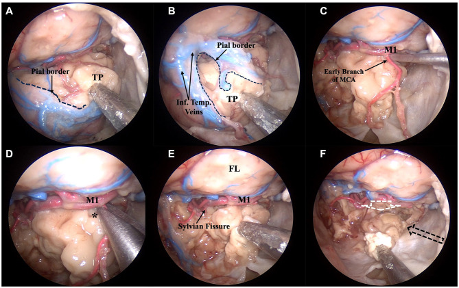
Anatomic pictures showing a right transorbital endoscopic approach for the resection of the temporal lobe. Steps 3 and 4, corresponding to the anterior corticectomy (A, B) and subpial dissection (C–F), respectively, are demonstrated. After the identification of the temporal pole and identification of all the surfaces of the temporal lobe, a small incision over the pia mater covering the temporal pole is made (A) and resection of the temporal lobe thus proceeds in a subpial plane (B). Mimicking what happens in a surgical scenario, the main vessels nourishing the temporal lobe are identified (C) and resected (D, *) before resection is continued (E). The direction of the resection parallels the sylvian fissure superiorly (white dotted arrow), the lateral wall of the cavernous sinus and tentorial incisura, medially (black dotted arrow), and the middle fossa floor inferiorly (direction given by the surgical aspirator) (F). * Early branch of the middle cerebral artery being dissected; FL, frontal lobe; Inf. Temp. Veins, inferior temporal veins; M1, sphenoid segment of the middle cerebral artery; TP, temporal pole.

