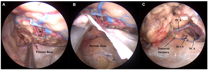
Figure 3.
Anatomic pictures showing a right endoscopic transorbital approach for the resection of the temporal lobe. Step 5, corresponding to the final exposure of the posterior landmarks identified after maximal resection of the temporal lobe, is represented. As the posterior limits of the temporal lobe are poorly represented and difficult to identify, we selected as the postero-lateral limit of our resection the petrous bone, whose position can be verified intradurally with a surgical instrument pointing at the petrous ridge (dotted line) (A). In (B), with an extradural approach to the middle cranial fossa, the dura mater is elevated in order to confirm our position (B). The postero-medial limit is represented by the junction between the crural (green dotted area) and interpeduncular cisterns (blue dotted area) (C). III CN, oculomotor nerve; M2, insular segment of middle cerebral artery; PCA, posterior cerebral artery; SCA, superior cerebellar artery.

