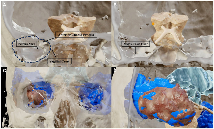
Figure 5.
BrainLab® reconstructions of post-dissection CT and MRI scans highlighting the relationship between the amount of bone removal (A, B) with the amount of resection of the temporal lobe (C, D). The bone work of the approach is limited to an anterior sphenoidal craniotomy (dark dotted area) which provides access to the temporal pole. Three of the four bony pillars of the approach, namely, the sagittal crest, the anterior clinoid process, and the petrous apex are left intact (A). The middle cranial fossa floor, which represents the fourth bony pillar, must be flattened to provide adequate maneuverability for the instruments which must work in parallel to the basal surface of the temporal lobe (B). The amount of removal of the temporal lobe (brown reconstruction) in relation with the total of the temporal lobe (blue reconstruction) is shown in an anterior (C) and antero-lateral (D) perspective.

