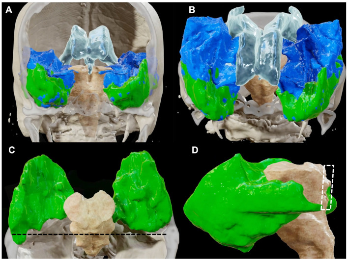
Figure 6.
BrainLab® reconstructions retrieved from post-dissection MRI quantifying the amount of temporal lobe removed (in green) in relation with the intact temporal lobe (in blue), seen from an anterior (A) and superior (B) perspective. In (C) and (D), only the temporal lobe removed was left to highlight the relationships of the posterior limits of the resection with the surrounding brain structures. The posterior limit of the resection is represented by a plane tangential to the lamina quadrigemina [dark dotted line in (C) and white dotted area in (D)] as it is demonstrated in a superior (C) and lateral (D) view. The qualitative analysis of our resection shows that while the middle and inferior temporal gyruses together with the basal and medial portion of the temporal lobe represented the largest amount of the resection, the posterior two-thirds of the superior temporal gyrus were left intact.

