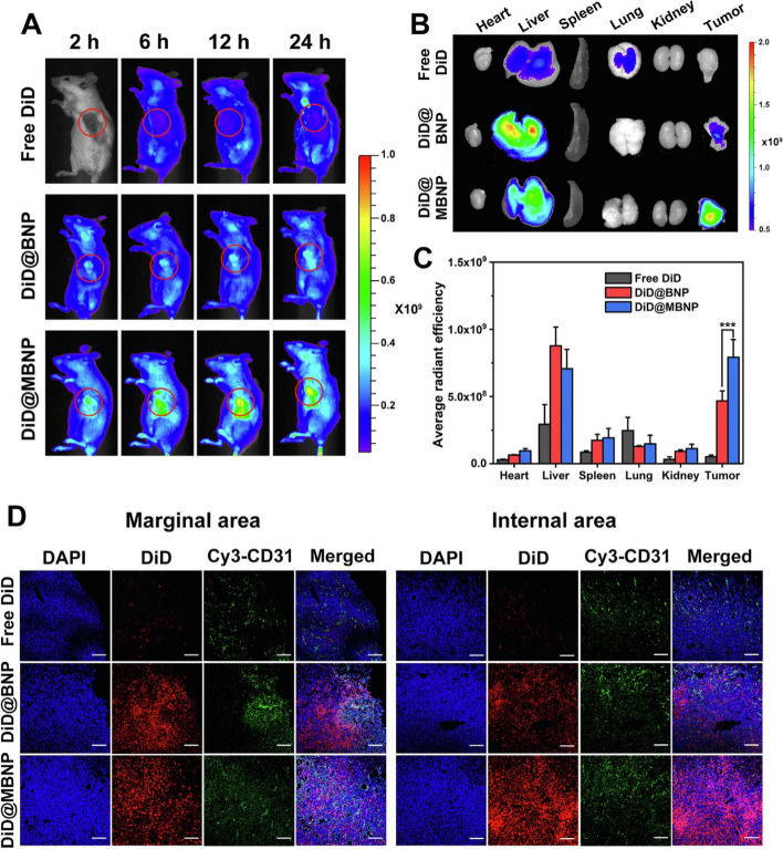
Fig. 3.
The results of the in vivo biodistribution of nanocarriers. The study involved the use of DiD-loaded formulations, and the images obtained from 4T1 tumor-bearing mice were taken at different times post-administration. The red circles in the images indicate the tumor sites. Additionally, ex vivo imaging of isolated tumors and organs from the mice was performed 24 h after administration. The semiquantification of fluorescence intensity was also done, and the results showed statistically significant differences (P < 0.05, P < 0.01, P < 0.001) among the three formulations. Finally, the fluorescent distribution of Free DiD, DiD@BNP, and DiD@MBNP in the frozen sections of tumors was analyzed, with blue indicating the cell nucleus, red indicating DiD, and green indicating CD31. The scale bars used in the images are 100 mm. Reprint from [111] with a permission from Elsevier

