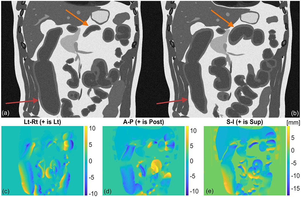
Figure 5.

Gastrointestinal motility in a simulated 4D T2-weighted MRI dataset demonstrating peristalsis and HAPC in the small and large bowel. Panel (a) and (b) show the wave at two separate timepoints. Arrows point to the location of the wave origin in each organ. Panels (c,d,e) show magnitude of displacement in the Lt-Rt, Ant-Post, and Sup-Inf direction, respectively. The deformation field relates displacement from motion state in panel (a) to motion state in panel (b).
