Figure 2.
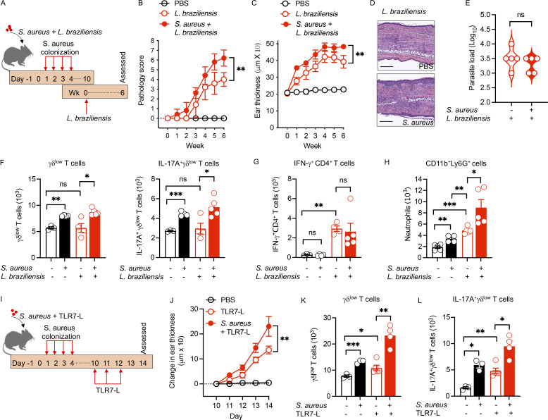
S. aureus augments pathology of cutaneous leishmaniasis and dermatitis. (A) Clinical S. aureus colonization and L. braziliensis infection protocol. C57BL/6 mice were colonized with S. aureus on days 1–4 and then on day 10 (week 0) mice were infected with L. braziliensis. Mice were euthanized at week 6 for tissue collection and analysis. (B and C) Pathology score (B) or ear thickness (C) of S. aureus–colonized or PBS control mice infected with L. braziliensis was assessed. Mean ± SD representative of two experiments with five mice per group. (D) H&E-stained ear sections of S. aureus–colonized or PBS control mice infected with L. braziliensis at week 6. Scale bar = 200 μm. (E) Parasite load in S. aureus–colonized or PBS control mice infected with L. braziliensis at week 6. Representative of two experiments with five mice per group. (F) Absolute numbers of γδlow T cells and IL-17A+γδlow T cells of S. aureus–colonized or PBS control mice infected or non-infected with L. braziliensis. Mean ± SEM representative of two experiments with three to five mice per group. (G) Absolute numbers of IFN-γ+CD4+ T cells of S. aureus–colonized or PBS control mice infected or non-infected with L. braziliensis. Mean ± SEM representative of two experiments with three to five mice per group. (H) Absolute numbers of CD11b+Ly6G+ cells of S. aureus–colonized or PBS control mice infected or non-infected with L. braziliensis. Mean ± SEM representative of two experiments with four to five mice per group. (I) S. aureus colonization and TLR7-L (imiquimod) treatment protocol. 6-wk-old C57BL/6 mice were colonized with S. aureus at days 1–4 and then treated with TLR7-L every day for 3 d (days 10, 11, and 12). Mice were euthanized on day 14 for tissue collection and analysis. (J) Skin thickness measurement of S. aureus–colonized or PBS control mice treated with TLR7-L. Mean ± SD representative of two experiments with five mice per group. (K and L) Absolute numbers of γδlow T cells and IL-17A+γδlow T cells of S. aureus–colonized or PBS control mice treated or non-treated with TLR7-L. Mean ± SEM representative of two experiments with three to five mice per group. *P < 0.05, **P < 0.01, and ***P < 0.001. “ns” denotes not significant. Significant P values by two-tailed unpaired Student’s t test with Welch’s correction are indicated in the figure.

