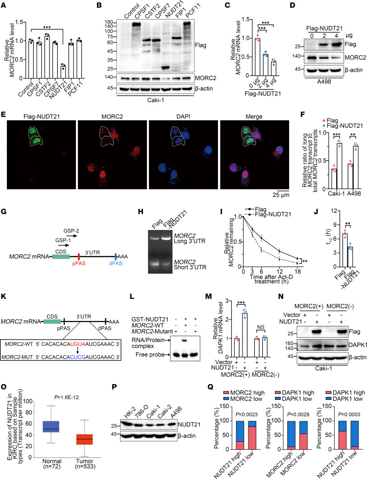
Figure 7. Loss of APA regulator NUDT21 induces 3′UTR shortening and upregulation of MORC2 in KIRC.
(A and B) qPCR (A) and immunoblots (B) were performed to evaluate MORC2 expression in Caki-1 cells transfected with indicated plasmid (n = 3). (C and D) qPCR (C) and immunoblots (D) were performed to evaluate MORC2 expression in A498 cells transfected with Flag vector or Flag-NUDT21 plasmid (n = 3). (E) Immunofluorescence staining was performed to evaluate MORC2 expression in Caki-1 cells transfected with Flag-NUDT21 plasmid. Scale bar: 25 μm. (F) qPCR was performed to evaluate the ratio of long 3′UTR MORC2 expression/total MORC2 expression in KIRC cells transfected with Flag vector or Flag-NUDT21 plasmid (n = 3). (G and H) 3′RACE was performed with GSP-1/2 primers (G) to evaluate 3′UTR of MORC2 in Caki-1 cells transfected with Flag vector or Flag-NUDT21 plasmid. (I and J) qPCR was performed to quantitatively analyze half-life of MORC2 in Caki-1 cells transfected with Flag vector or Flag-NUDT21 plasmid (n = 3). (K) Diagram indicated the UGUA motif of MORC2 and the mutant motif. (L) EMSA was performed to explore the binding site of NUDT21 at MORC2 3′UTR. (M and N) qPCR (M) and immunoblots (N) were performed to evaluate DAPK1 expression in WT and MORC2-depleted Caki-1 cells transfected with Flag vector or Flag-NUDT21 plasmid (n = 3). (O) NUDT21 expression in KIRC tissues and normal kidney tissues was analyzed with UALCAN database. (P) Immunoblotting was performed to evaluate NUDT21 expression in HK-2 cells and KIRC cells. (Q) The expression associations between NUDT21 and MORC2/DAPK1, and between MORC2 and DAPK1, were analyzed in KIRC specimens. All data represent the mean ± SD. Two-tailed t test analyses were performed. **P < 0.01; ***P < 0.001.

