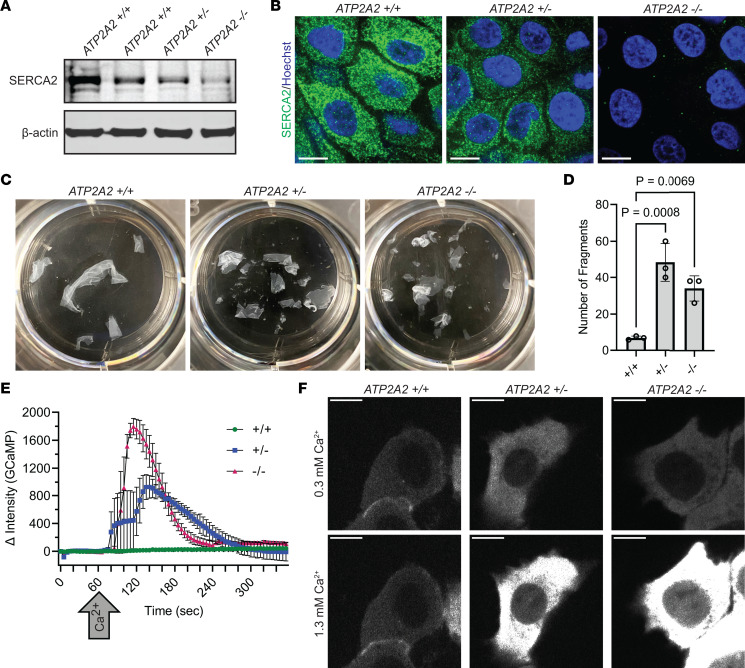
Figure 1. Loss of SERCA2 in human keratinocytes impairs cytosolic calcium handling and reduces intercellular adhesive strength.
(A) Immunoblot of SERCA2 in lysates from ATP2A2 wild-type (WT, +/+), heterozygous (HET, +/-), and homozygous knockout (KO, -/-) cells. hTERT-immortalized human epidermal keratinocytes (THEKs) were differentiated in E-medium for 72 hours before lysate harvesting; data represent 3 independent experiments; and β-actin is a loading control. (B) Immunofluorescence of SERCA2 (green) in WT, HET, and KO THEKs; images are representative of 14 independent high-powered fields (hpf) per genotype; Hoechst (blue) stains nuclei; scale bar = 10 μm. (C) Mechanical dissociation of monolayers from control (+/+), HET (+/-), and KO (-/-) cells grown in 1.3 mM CaCl2 for 72 hours prior to using dispase to release intact monolayers; representative images of fragmented monolayers transferred into 6-well cell culture plates after mechanical stress are shown. (D) Graphs display mean ± SD of the number of epithelial fragments with data plotted for N = 3 biological replicates; P values from 1-way ANOVA with Dunnett’s adjustment for multiple comparisons. (E) Change (Δ) in intensity of GCaMP in control (+/+), HET (+/-), and KO (-/-) THEKs from baseline at t = 0 seconds with addition of 1 mM CaCl2 at t = 60 seconds (arrow); data plotted as mean ± SEM from N = 3 independent experiments per genotype. (F) Representative fluorescence images of GCaMP in WT, HET, and KO cells in low CaCl2 (left) or high CaCl2 (right); scale bar = 10 μm.

