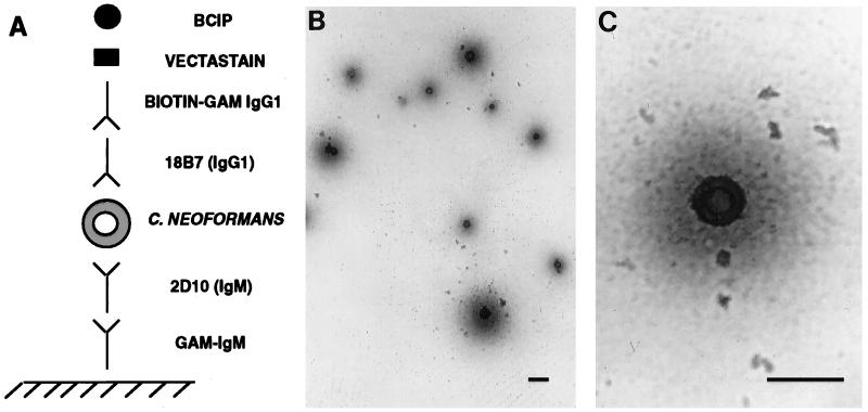
FIG. 2.
Spot ELISA for C. neoformans with MAb 18B7. (A) Graphic representation of the ELISA configuration. GAM, goat anti-mouse. (B and C) Light microscopy images of C. neoformans 24067 captured and detected by the assay. In this assay the C. neoformans cells stain blue. Magnification, ×200 (B) and ×400 (C). Bars, 20 μm.

