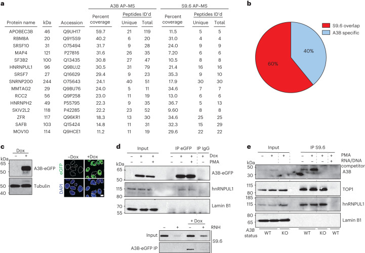
Fig. 1. APOBEC3B (A3B) interacts with R-loop-associated proteins.
a,b, Shared proteins in A3B and S9.6 AP–MS datasets. c, Immunoblot and IF microscopy analysis of MCF10A-TREx-A3B-eGFP cells treated with vehicle or Dox (1 µg ml−1, 24 h). A3B-eGFP (green) is predominantly nuclear (DAPI, blue). Ten-micrometer scale bar; n = 2 (left); n = 1 (right) biologically independent experiments. d, Immunoblots of indicated proteins in A3B-eGFP or IgG IP from TREx-A3B-eGFP MCF10A cells ± Dox (1 μg ml−1, 24 h), treated with PMA (25 ng ml−1, 2 h) and probed with indicated antibodies (top). Slot blot of A3B-eGFP IP from TREx-A3B-eGFP MCF10A cells ± Dox (1 μg ml−1, 24 h) ± exogenous RNase H (RNH) probed with S9.6 antibody (bottom). n = 2 biologically independent experiments. e, Immunoblots of indicated proteins in S9.6 IP reactions from MCF10A WT or A3B KO cells treated with PMA (25 ng ml−1, 5 h). n = 2 biologically independent experiments.

