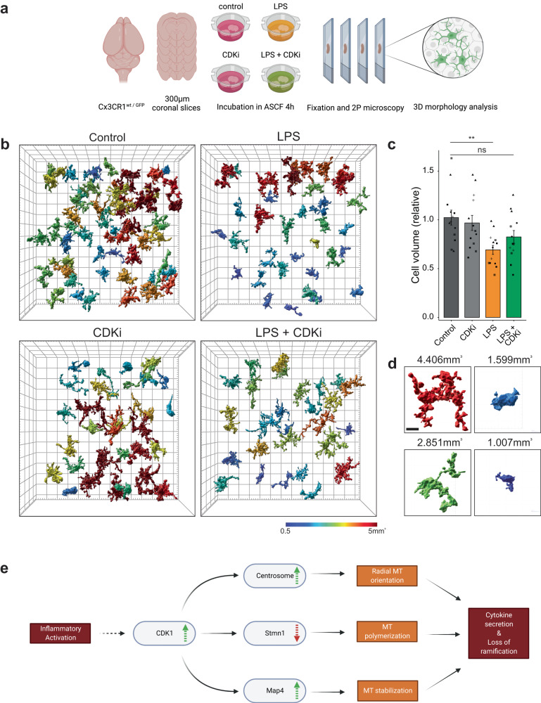
Fig. 7. Cdk1 inhibition prevents morphological remodeling of LPS-reactive microglia in ex-vivo brain tissue.
a Experimental outline: 300 µm thick acute slices of Cx3CR1wt/GFP brains were incubated in treatments indicated in ASCF for 4 h before fixation and 2-photon microscopy. b Representative 3D segmentation result of GFP signal from 150 µm thick image stacks of indicated treatments. Cells are color coded by their volume. Grid spacing is 20 µm in xy and 40 µm in z. See also Supplementary Video 3. c Quantification of cell volumes relative to control condition in acute slices shown in (b). Graph shows mean ± SE for n = 13 acute sections recorded in 3 ROIs each from 5 mice. Symbols indicate datapoints originating from the same animals. Statistical significance was calculated with ANOVA and Tukey HSD. Source data are provided as a Source Data file. Significance intervals p: **** <1e−04 <*** <0.001 <** <0.01 <* <0.05 <ns. d Example 3D segmentation of cells from b with measured volumes indicated. Color-coding as in (b). Scale bar is 5 µm. e Working model of microglial inflammatory stimulation and resulting MT reorganization as proposed in this paper.

