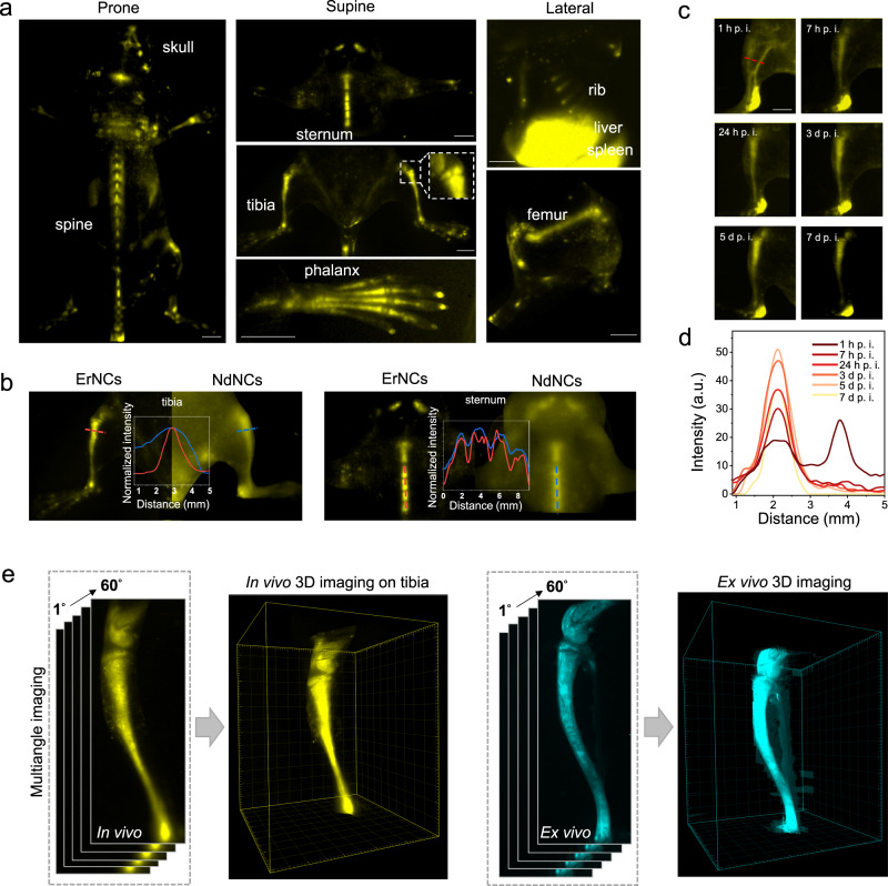
Fig. 1. High-resolution NIR-II imaging of mice bones in both two and three dimensions.
a NIR-II in vivo imaging of murine bones in prone (skull, spine), supine (sternum, tibia, phalanx, subchondral bone in the zoom-in picture), and lateral (rib, femur) postures by the 1550 nm fluorescent ErNCs (980 nm excitation, 38 mWcm−2, 1319 nm long-pass). Scale bars: 2 mm. b The NIR-II imaging of bone and corresponding cross-sectional intensity profiles by ErNCs (980 nm excitation, 38 mWcm−2, 1319 nm long-pass) and NdNCs (808 nm excitation, 34 mWcm−2, 900 nm long-pass), respectively. c In vivo NIR-II imaging of mouse tibia by ErNCs (980 nm excitation, 38 mWcm−2, 1319 nm long-pass, 60 ms) at the different post-injection time points (d: day). Scale bar: 2 mm. d Cross-sectional intensity profiles along the same position of the time series images represented by the red dash line in c. e The 3D NIR-II imaging on mouse tibia reconstructed by a series of images recorded under different angles (0°−60°) rotated along the central axis of the tibia.

