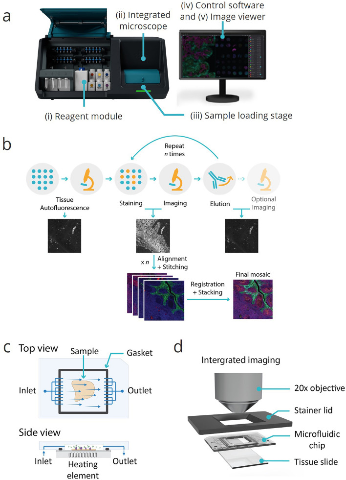
Figure 1.
Overviews of COMET™ platform and seqIF method, enabling full automation and walk-away execution of multiplex immunofluorescence assays. (a) COMET™ platform overview. (i) A reagent module composed of 20 small volume reservoirs for reagents, typically used for primary antibodies, 4 mid-size reservoirs, typically used for detection reagents, and 7 large volume reservoirs for ancillary buffers; (ii) an integrated epifluorescence microscope acquiring automated mosaic scans of stained samples, (iii) a sample loading/unloading interface with a rotary stage that accommodates 4 automated stainers, (iv) a control software for protocol preparation and execution; and (v) an imaging viewer for rapid visualization of multiplexed immunofluorescence results. (b) Sequential immunofluorescence method (seqIF). Tissue autofluorescence is automatically acquired as a baseline for downstream background removal. Samples are automatically stained and imaged for DAPI and 2 markers of interest. Elution—signal removal by chemical removal of antibodies. Staining, imaging, and elution are repeated n times (n: number of multiplexed staining steps). Images are automatically stitched, aligned, and stacked on a final OME-TIFF file. (c) Top and side views of the microfluidic imaging chip consisting of inlet, outlet and a fluidic network that enable uniform exposure of reagents over the tissue sample. A gasket at the edges of the staining area ensures a hermetic sealing. A heating element underneath is used to control the temperature of each step in the seqIF protocol. (d) Integrated imaging assembly. The microfluidic chip contains an imaging window that allows the integration of in-situ fluorescence microscopy and permits the direct imaging of the sample.

