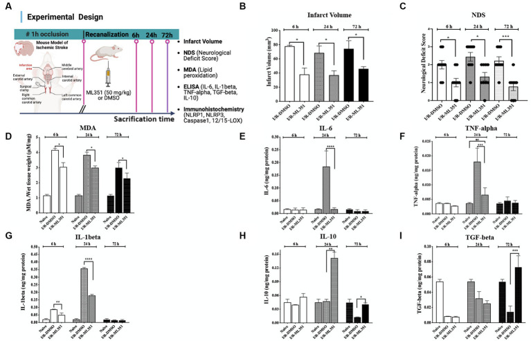
Figure 1.
Determination of infarct volume, neurological deficit, lipid peroxidation, and pro- and anti-inflammatory cytokine levels in the brain tissues of I/R-DMSO and I/R-ML351 mice. (A) The experimental design of the study is summarized in the schema. Briefly, ischemia/recanalization (I/R) was performed by proximal middle cerebral artery occlusion in mice. Either the 12/15-LOX inhibitor (ML351, 50 mg/kg) or its solvent, DMSO, was injected i.p. at recanalization after 1-h of occlusion. Mice were sacrificed at 6, 24, and 72-h after ischemia induction. (B) Infarct volume analysis was performed on Nissl-stained sections. Infarct volumes were significantly decreased in ML351-treated groups at 6, 24, and 72-h (n = 3 mice/group, *p ≤ 0.05, *p = 0.021, *p ≤ 0.050, respectively). (C) Functional outcome was determined with neurological deficit scoring following I/R. Neurological deficit score results showed that 12/15-LOX inhibition significantly improved neurological deficit at all time points (n = 9 mice/group, *p = 0.0420 for 6-h; *p = 0.0170 for 24-h; ***p = 0.0008 for 72-h). (D) MDA analysis results showed that increased lipid peroxidation after I/R was attenuated with ML351-treatment at all time points (n = 3 mice/group, ***p = 0.0004 for 6-h, *p = 0.0196 for 24-h, *p = 0.0414 for 72-h). (E–G) Quantitative analysis of IL-6, TNF-alpha, and IL-1beta pro-inflammatory cytokines was done with ELISA (n = 3 mice/group), data is given as proportioned to the total protein levels of the brain tissue samples (ng/mg protein). Pro-inflammatory cytokines were induced especially at 24-h following I/R. IL-6 (****p < 0.0001) and TNF-alpha (***p < 0.0004) were suppressed by administration of ML351 at 24-h of ischemia. IL-1beta was increased at 6-h (***p = 0.0002) and 24-h (****p < 0.0001) of I/R and decreased with ML351 treatment at 6-h (**p = 0.003) and 24-h (****p < 0.0001). (H,I) Quantitative analysis of anti-inflammatory cytokines, IL-10, and TGF-beta, with ELISA (n = 3 mice/group). ML351 induced the IL-10 anti-inflammatory cytokine at 24-h and 72-h of I/R (**p = 0.0069, *p = 0.0192, respectively), while TGF-beta was increased at 72-h of I/R by ML351 treatment (***p = 0.0002).

