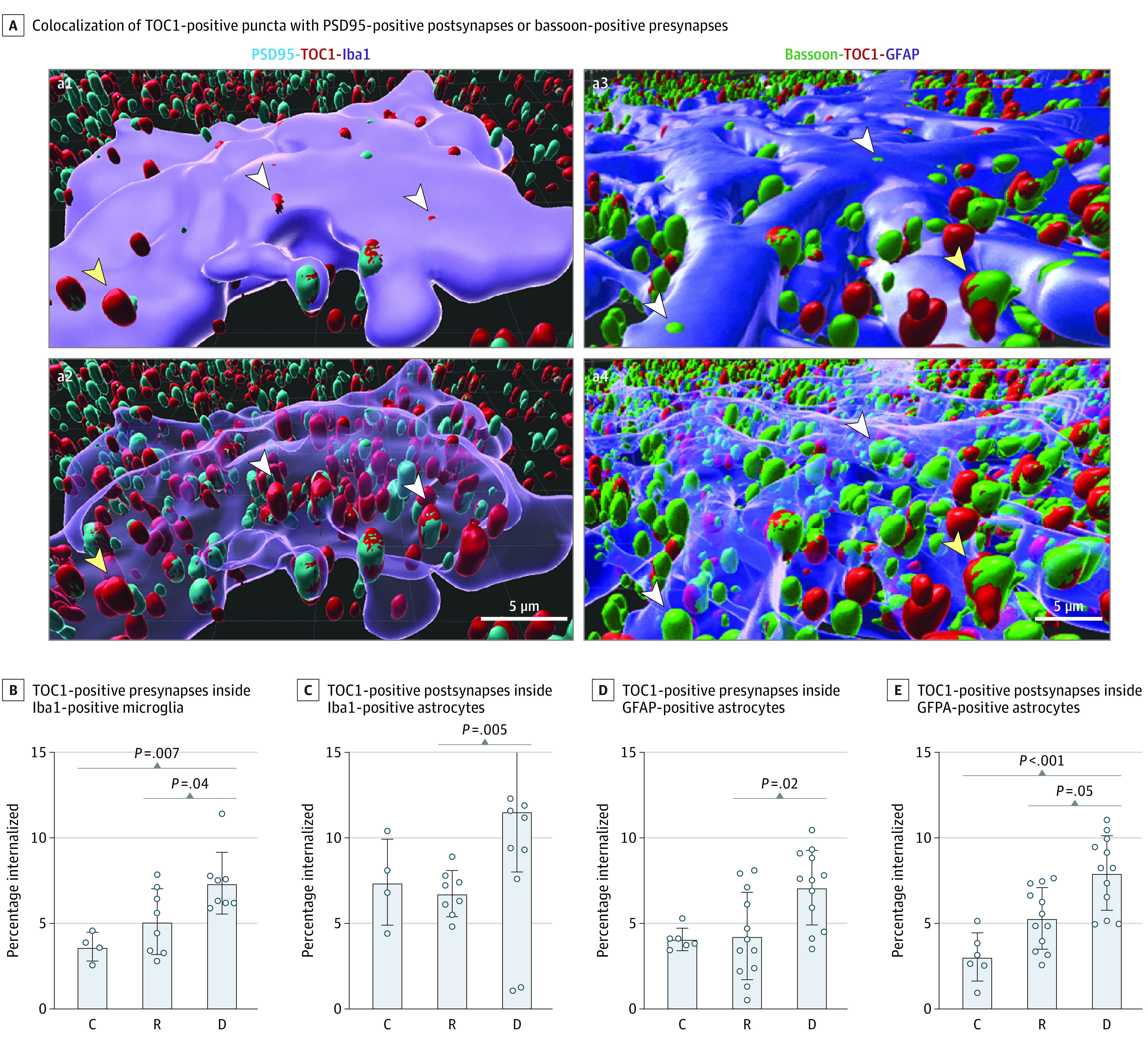
Figure 4. Glial-Mediated Engulfment of Tau Oligomer–Tagged Synapses.

A, Representative 3-dimensional Imaris reconstructed images of postsynaptic density 95 (PSD95)–positive with tau oligomeric complex 1 (TOC1)–positive synapses inside an ionized calcium-binding adaptor protein molecule 1 (Iba1)–positive ameboid microglia before (a1) and after (a2) making the cell body transparent, and bassoon-positive with TOC1-positive synapses inside a glial fibrillary acidic protein (GFAP)–positive astrocyte before (a3) and after (a4) making the cell body transparent. White arrowheads indicate engulfed; yellow arrowheads, not engulfed. Assessments of percentages of internalized TOC1-positive presynapses (B) and TOC1-positive postsynapses (C) inside Iba1-positive ameboid microglia and GFAP-positive astrocytes (D and E). Analyses were performed for 20 ionized calcium-binding adaptor molecule 1–positive and 30 GFAP-positive cells from 10 brains (2 controls, 4 resilient, 4 dementia). C indicates control (Braak stage 0-II); D, dementia (Braak stage III-IV); and R, resilient (Braak stage III-IV).
