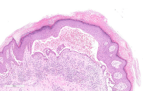
Figure 2.

Histological examination of the skin punch biopsy showed suprabasal blister formation and acantholysis. The blister cavity contained acantholytic cells, eosinophils, and neutrophils. There was a moderate perivascular inflammatory infiltrate composed of lymphocytes, plasma cells, neutrophils, and eosinophils (hematoxylin-eosin stain, magnification ×10).
