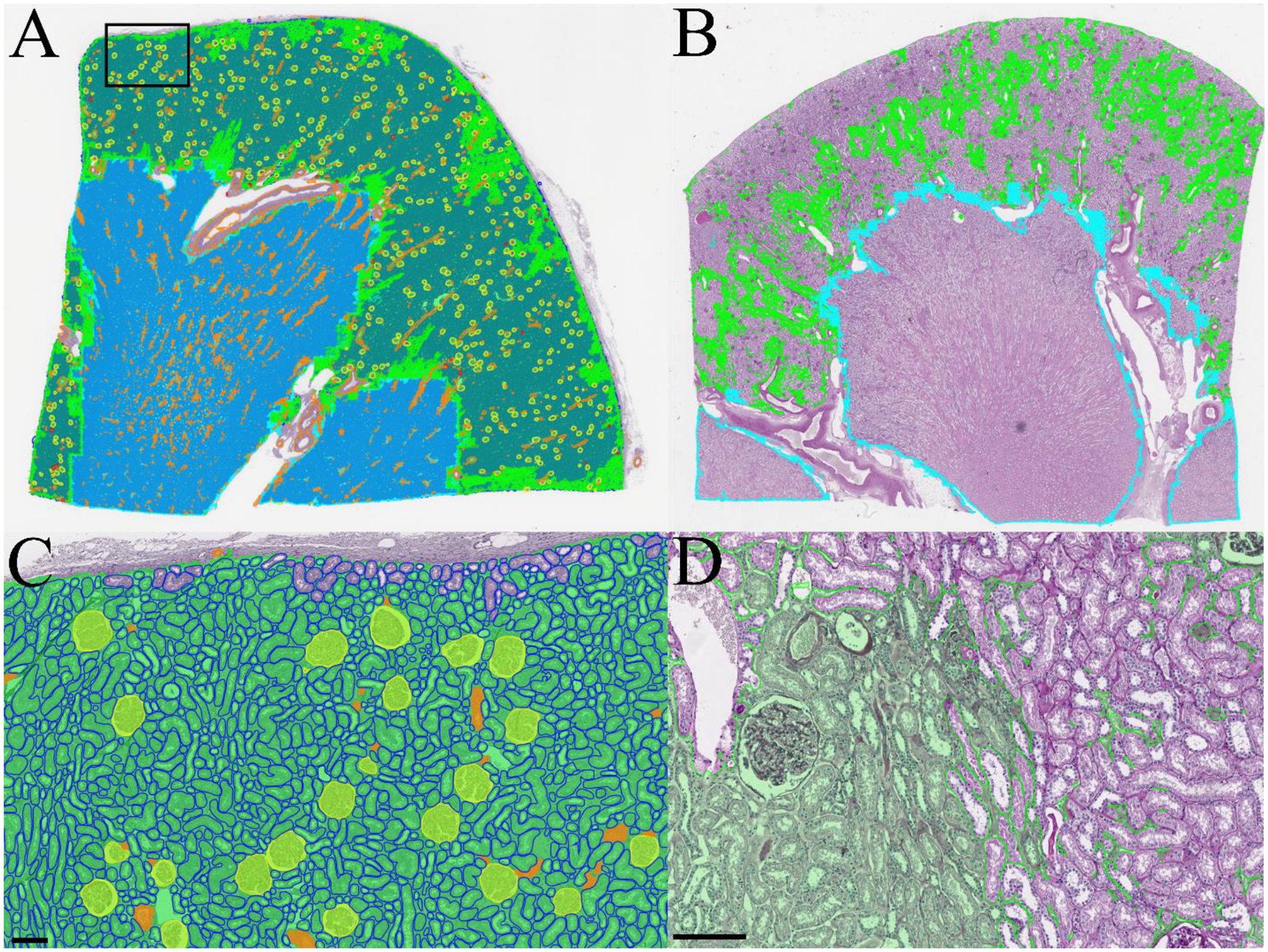
Figure 1.

Whole section segmentations for PAS-stained kidney nephrectomies. A) Thumbnail of a whole segmentation mask for a reference kidney. Tubules are rendered in the background to prevent them from overwhelming the visibility of other structures. B) Thumbnail of patchy interstitial segmentation in a kidney with many tubules flush back to back. C) Zoomed region from A showing segmentation of viable glomeruli, tubules, arterioles, and cortical interstitium. D) Zoomed region from B showing interstitium at left fused by contour retrieval after tile stitching process, where interstitium at right is patchy due to flushly abutting tubules. Green: cortical interstitium; cyan: medullary interstitium; yellow: viable glomerulus; red: sclerotic glomerulus; blue: tubule; and orange: artery/arteriole. Scale bar 150μm.
