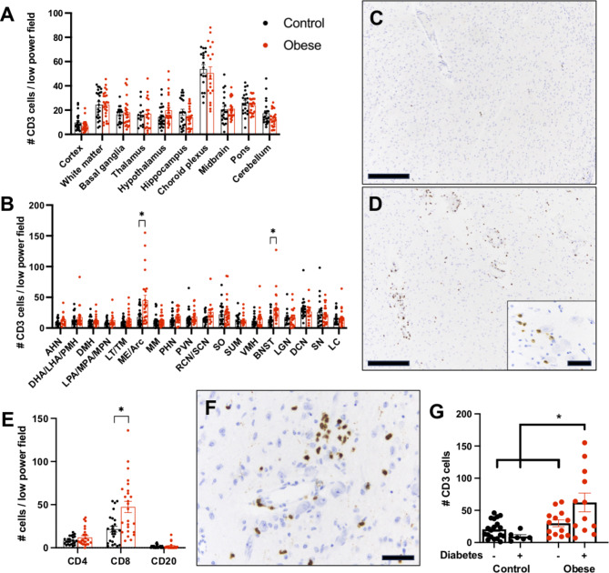
Fig. 1.
T cell infiltrates are increased in hypothalamic arcuate/medial eminence and in bed nucleus of the stria terminalis of obese humans. CD3-positive cells were quantified in standardized post-mortem brain tissue sections from control and obese humans (A). CD3-positive T-cells in various hypothalamic regions and other non-hypothalamic brain nuclei from control and obese humans (B). Representative CD3 immunohistochemistry in the Arc/ME of a non-obese (C) and obese (D) individual. Note the close apposition of CD3-positive T-cells to hypothalamic neurons (D, inset). Quantification of CD4, CD8, and CD20 inflammatory cells in the Arc/ME from control and obese individuals (E). Representative CD8 immunohistochemistry from the Arc/ME from an obese human (F). CD3-positive cells in the Arc/ME of non-obese/obese individuals with and without a clinical history of diabetes (G). Scale bars: A,D = 500 μm, D(inset), F = 50 μm. * p-value < 0.05

