. 2023 Jan 13;7(1):1–16. doi: 10.4049/immunohorizons.2200091

FIGURE 4.

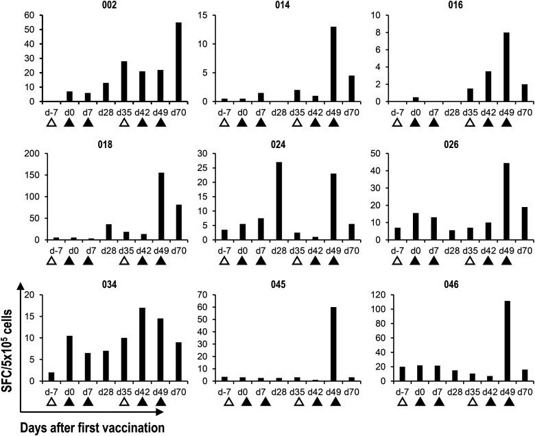
FIGURE 4. Detection of α-GalCer–reactive IFN-γ–producing cells by the ELISPOT assay. Cryopreserved PBMCs were thawed and cultured for 16 h in the presence of α-GalCer. IFN-γ–producing cells were analyzed by an IFN-γ ELISPOT assay. The mean IFN-γ spot-forming cell (SFC) number for duplicate cultures in nine representative cases is shown. Apheresis (△) and APC/Gal (▲) are shown.

Detection of α-GalCer–reactive IFN-γ–producing cells by the ELISPOT assay.

Cryopreserved PBMCs were thawed and cultured for 16 h in the presence of α-GalCer. IFN-γ–producing cells were analyzed by an IFN-γ ELISPOT assay. The mean IFN-γ spot-forming cell (SFC) number for duplicate cultures in nine representative cases is shown. Apheresis (△) and APC/Gal (▲) are shown.