
Figure 5. Treatment of mice with miRNA 130b‐3p mimic attenuates eCIRP‐induced pro‐inflammatory responses and improves overall lung histology.
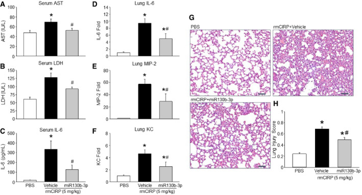
Mice were injected with PBS or rmCIRP (5 mg/kg BW) i.v. together with or without miRNA 130b‐3p at a dose of 12.5 μl of 1,000 μM. In the miRNA 130b‐3p treatment group, rmCIRP and miRNA 130b‐3p mimic were combined 30 min prior to administration into mice intravenously.
-
A–FAfter 5 h of rmCIRP or miRNA 130b‐3p injection into mice, blood samples and lung tissues were collected to assess (A) AST, (B) LDH, (C) and IL‐6 in serum and mRNA for (D) IL‐6, (E) MIP‐2, and (F) KC in the lungs. AST, aspartate aminotransferase; LDH, lactate dehydrogenase; MIP‐2, macrophage inflammatory protein‐2; KC, keratinocyte chemoattractant.
-
GLung tissue was collected after 5 h from PBS‐ and rmCIRP (5 mg/kg BW)‐treated mice with or without miRNA 130b‐3p mimic (12.5 μl of 1,000 μM) and stained with H&E. Each slide was observed under light microscopy at 400× original magnification. Representative images for each group are shown. Scale bar, 50 μm.
-
HHistological injury scores of the lungs in different groups were quantified as described in Materials and Methods.