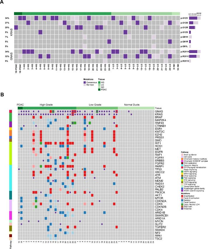
FIGURE 1.
Genomic landscapes of IPMN lesions. A, SNVs identified in KRAS and GNAS in LG and HG IPMNs. Dark purple: somatic mutations detected for KRAS and GNAS. Light purple: mutations detected by retesting. B, SNVs and CNAs identified in ND, LG, HG, and PDAC regions and classified by relevant PDAC-related pathways. For A and B, samples were arranged by histological type and labeled at the top of each heat map.

