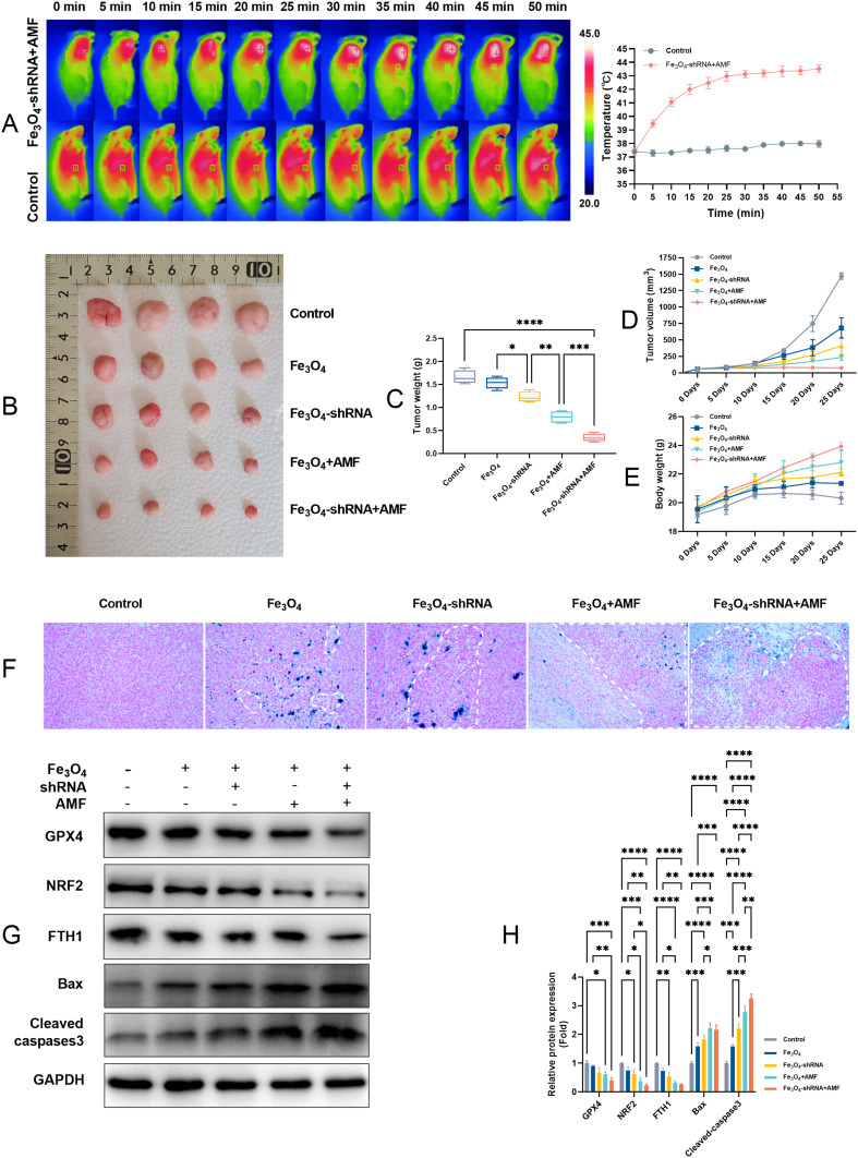
Figure 6.
The in vivo therapeutic effects of the MUC1-C shRNA@Fe3O4 MNPs on TNBC. IR thermal images of TNBC-bearing C-NKG mice with local injection of MUC1-C shRNA@Fe3O4 MNPs (200 µg mL−1), or normal saline under AMF (3Kw) (A). Images (B) and weights (C) of xenograft tumors harvested from the mice after various treatments on the 25th day. Tumor volumes (D) and Body weight (E) growth in mice were measured after different treatments every 5 days within 25 days (n=4). Prussian blue reaction of tumor tissue showing iron staining and tumor necrosis, labeled area was typical necrosis (200x) (F). Expression of Bax, cleaved-caspase 3, GPX4, NRF2, and FTH1 in tumor tissues (G). Quantitative statistical results according to the protein bands (H). Error bars represent means ± SD. *p<0.05, **p<0.01, ***p<0.005, ****p<0.001.

