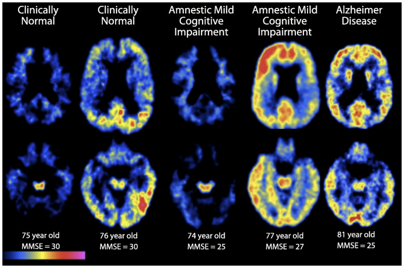
Figure 1-1.

Carbon-11 Pittsburgh Compound B positron emission tomography (PET) amyloid images show two transaxial slice levels in six older individuals, with age and Mini-Mental State Examination (MMSE) score listed at the bottom. Regions of red and yellow indicate high Pittsburgh Compound B retention, indicating presence of fibrillar amyloid deposition. From left to right: a 75-year-old clinically normal woman who is amyloid negative; a 76-year-old clinically normal man with evidence of amyloid deposition in parietal and temporal cortices; a 74-year-old woman with amnestic mild cognitive impairment (aMCI) and an MMSE of 25 who is amyloid negative; a 77-year-old man with aMCI and an MMSE of 27 demonstrating amyloid deposition in frontal, temporal, and parietal cortices; and an 81-year-old woman with mild Alzheimer disease dementia with an MMSE of 25 who demonstrates evidence of amyloid deposition in frontal, temporal, and parietal cortices.
