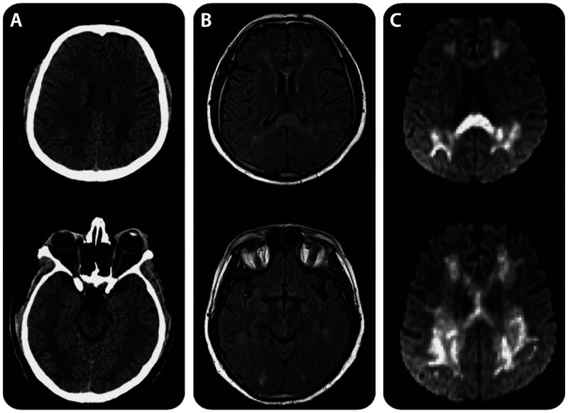
Figure 7-3.

CT and MRI features of Marchiafava-Bignami disease. A, Axial CT showing symmetric hypodense white matter lesions. B, Fluid-attenuated inversion recovery (FLAIR)–weighted MRI depicting hyperintense white matter lesions predominantly involving the splenium of the corpus callosum. C, Diffusion-weighted images revealing marked restriction with corresponding low apparent diffusion coefficient values. The mammillary bodies and the periaqueductal region appear normal. Reprinted with permission from Tozakidou M, et al, Neurology.16 © 2011, American Academy of Neurology. www.neurology.org/content/77/11/e67.long.
