Figure 7-2.

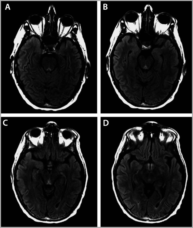
Brain MRI of a patient with Wernicke syndrome, as detailed in Case 7-2. MRI reveals T2 fluid-attenuated inversion recovery (FLAIR) hyperintensities in the periaqueductal gray (A, B), midbrain tectum (C), and mammillary bodies (D).

Brain MRI of a patient with Wernicke syndrome, as detailed in Case 7-2. MRI reveals T2 fluid-attenuated inversion recovery (FLAIR) hyperintensities in the periaqueductal gray (A, B), midbrain tectum (C), and mammillary bodies (D).