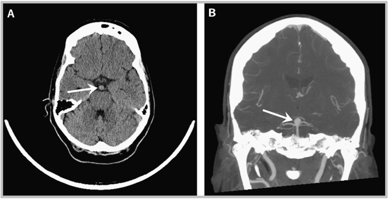
Figure 8-1.

Imaging studies taken of the patient in Case 8-1 at presentation to the emergency department. A, Noncontrast CT of the brain showing enlargement of the basilar artery near the bifurcation (arrow). B, CT angiogram (coronal view) showing basilar apex aneurysm (arrow).
