
Figure 3-16.
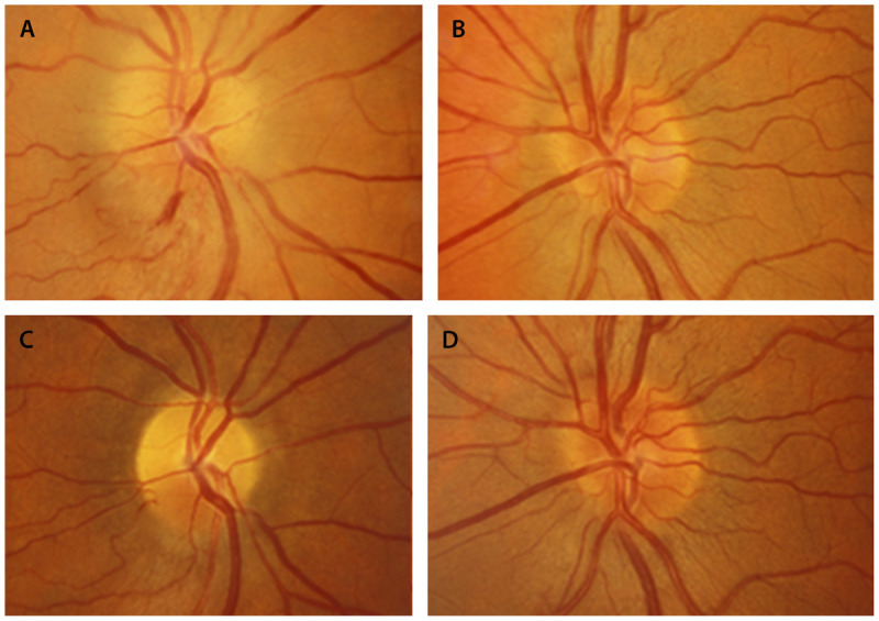
Nonarteritic anterior ischemic optic neuropathy in the right eye. A (right), B (left), Optic nerves 1 week after visual loss in the right eye. The right optic nerve is swollen with a small peripapillary hemorrhage. The left eye has a disc at risk for nonarteritic anterior ischemic optic neuropathy, with a small cup-disc ratio. C (right), D (left), Optic nerves 8 weeks later. In the right eye, the swelling has resolved, and segmental superior optic nerve head pallor is present in the right eye.
Reprinted with permission from Biousse V, Newman NJ, Thieme.1 © 2009 Thieme Medical Publishers, Inc.