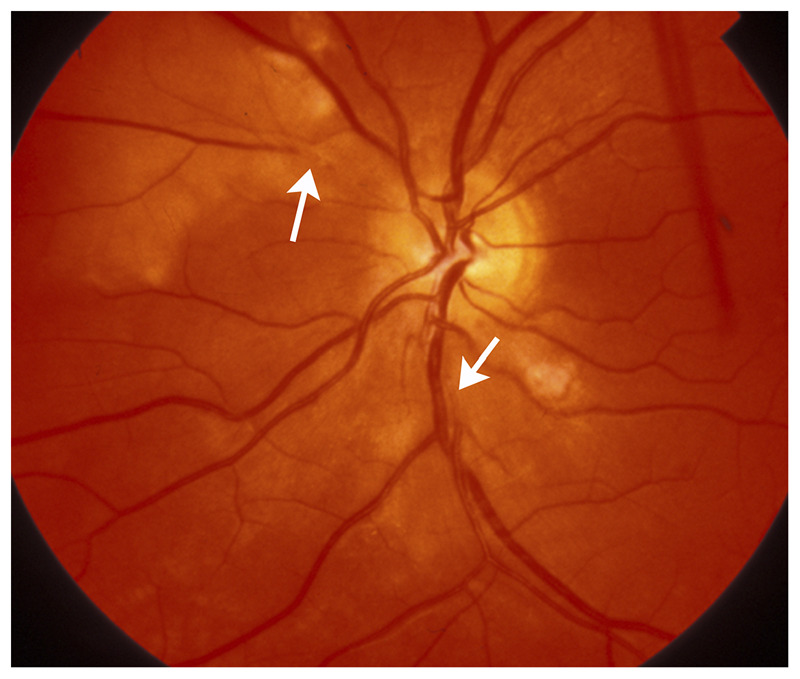
Figure 3-6.

Platelet-fibrin retinal emboli from aortic arch atheroma. Multiple grayish emboli (arrows) are seen in a few branches of the central retinal artery. The whitish areas in the retina correspond to associated retinal ischemia. Although the patient was not aware of visual loss, a visual field test showed multiple small scotomas corresponding to the areas of ischemic retina.
Reprinted with permission from Biousse V, Newman NJ, Thieme.1 © 2009 Thieme Medical Publishers, Inc.
