
Figure 2-12.
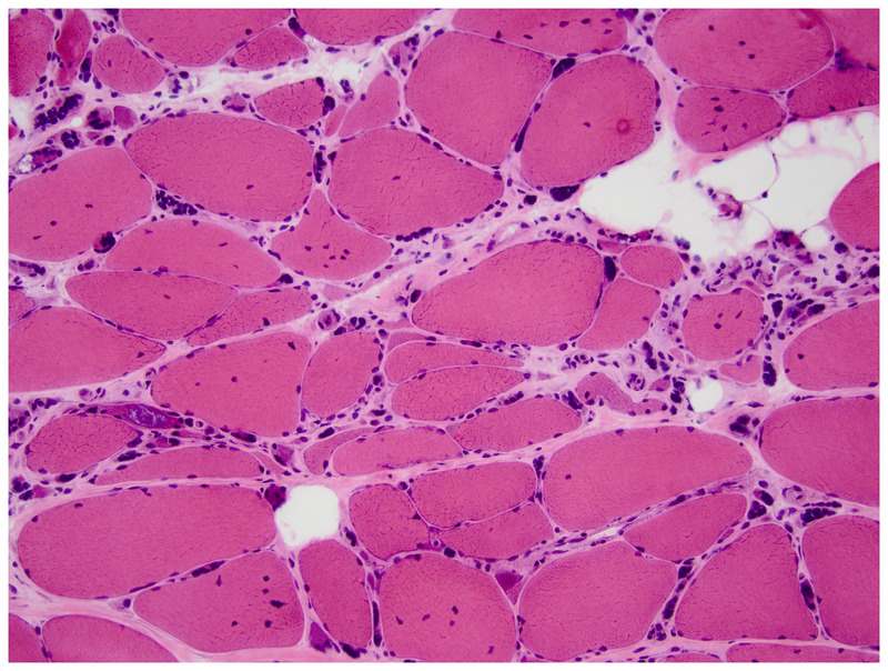
Muscle biopsy of a patient with myotonic dystrophy type 2. Note several muscle fibers with >5 internal nuclei and multiple pyknotic nuclear clumps superimposed on dystrophic appearing muscle (hematoxylin and eosin). Courtesy of Jennifer W. Baccon, MD, PhD.